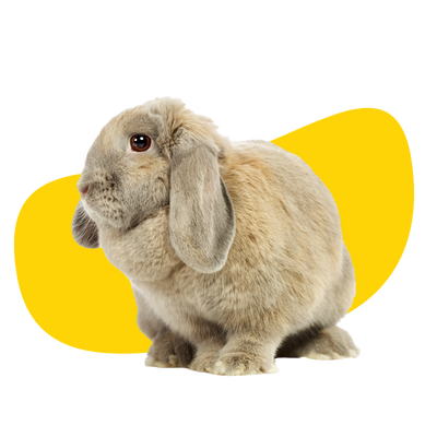
Lop-eared rabbits are highly susceptible to ear diseases, making ear surgery a common necessity. A summary of the most common clinical issues, diagnostic steps and surgical intervention is included below.
Clinical Issues
In lop-eared rabbits, the external ear canal is both kinked and narrowed (stenotic), which hinders the drainage of ear discharge. This creates a warm, moist environment that is ideal for infections to develop.
An external ear infection occurs when discharge accumulates in the external ear canal while the eardrum (tympanic membrane) remains intact, and the middle ear appears normal. Many rabbits show no obvious symptoms, but some signs to watch for include head shaking, ear scratching, hair loss around the base of the ears, and swelling of the ear canals. In some cases, the only noticeable changes might be a reduced appetite or episodes of recurrent gut stasis.
In some cases, ear infections can spread to the middle ear, affecting the tympanic cavity within the skull. This area, which is normally air-filled, becomes filled with pus during an infection. If the infection progresses further, it may also impact the brain structures responsible for balance (the vestibular system). While rabbits are incredibly stoic and may not show symptoms even at this advanced stage, signs can include mouth asymmetry, head tilt, unsteadiness, circling, and, in severe cases, seizures.
Medical management is often attempted, and some rabbits with external ear infections may experience temporary improvement. However, due to the unique anatomy of lop-eared rabbits, these infections frequently relapse. When the infection involves the middle ear, it is highly unlikely to resolve through medical treatment alone.
Imaging
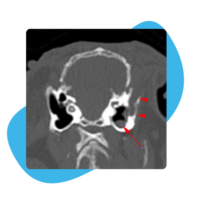
To accurately assess the severity of ear disease in your rabbit, we recommend imaging, with CT scans being the preferred method. CT scans provide a detailed view of the external ear canal and help determine if the infection has progressed to the tympanic cavity (middle ear). This allows us to develop a precise diagnosis and tailored treatment plan.
To the right is a CT image of a rabbit with otitis media (middle ear infection). The right ear has pus present in the external ear canal (red arrowheads) as well as in the tympanic cavity (red arrow), indicating a middle ear infection is present. The left ear has a tympanic cavity filled with air, showing up as black on the image, indicating a middle ear infection is not present.
Surgery
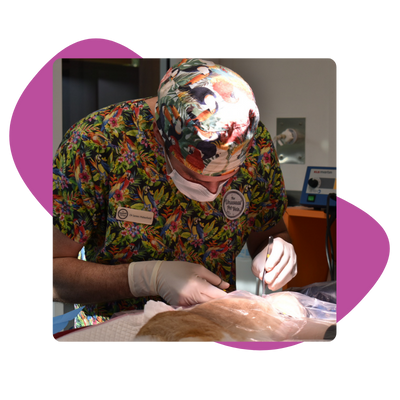
If your rabbit has an external ear infection (otitis externa), a lateral ear canal resection (LECR) is typically performed. This procedure involves removing part of the external ear canal and stitching it to the surrounding skin to create an open pathway for discharge to drain. While this surgery cannot completely prevent the development of a middle ear infection, it significantly reduces the likelihood.
If your rabbit has a middle ear infection (otitis media), the treatment involves both a LECR and either a bulla osteotomy (BO) or a myringotomy. During this procedure, the tympanic cavity of the skull is accessed, and the accumulated pus is carefully removed. The skin around the bulla is then sutured open, exposing the cavity to air and allowing for post-operative flushing (if needed) to ensure thorough cleaning and drainage.
The ideal outcome of the surgery is to allow the ear canal to have access to air and allow any discharge to drain. This improves comfort, and decreases infection risk, as well as making infections easier to treat. Samples for culture and sensitivity will usually be taken during surgery. This checks what bacteria are causing the ear infection, and what antibiotic will be most effective. Your rabbit will be sent home on antibiotics after surgery, but these may need to be changed depending on culture results.
Post-Operative Care
Because the surgery aims to open an area that was previously not open, the appearance of the ear after surgery can sometimes be quite confronting for owners, and there is commonly a lot of discharge and ooze from the surgical site. This needs to be cleaned daily to prevent it getting stuck in the stitches. If too much ooze collects then the surgical site can close over, as rabbits heal quickly. We will show you how to clean the discharge safely. If you are struggling to clean the area, please contact us so our staff can help you with this.
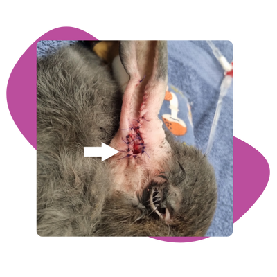
The image to the right (grey bunny) shows how the surgical site will look immediately after the surgery. The stoma (white arrow) is the very end of the ear canal that needs to remain open. It is important to keep the stoma as clean and free of discharge as possible. Any ooze collecting in the stitches should be cleaned at least daily. It is common for areas of the surgical site to break down (edges come apart), as we can’t really protect the bunny from cleaning or scratching the area. These areas will normally scab over with time.
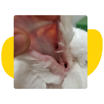
After your rabbit’s surgery, we will generally be seeing them once to twice per week until fully healed. This normally takes two to three weeks. Some rabbits with severe disease or particularly nasty bacteria may take longer to recover or occasionally require follow-up procedures.
Some clinical signs, such as a head tilt or facial asymmetry, may take longer to improve. In some cases, they will resolve, however some nerves are too badly damaged to heal so changes may be permanent, but the source of pain has been removed.
The image to the right (white bunny) shows how the surgical site generally looks after it’s completely healed, and the stitches are removed. The ear canal is open and can air out, and the hair is growing back. Any discharge or wax that the ear canal produces can now drain easily. When the ear is in a normal position (flopped), the surgical site is not visible. It will only be seen once the ear is lifted.

